
By Jill Gregory and Courtney McKenna
Mount Sinai’s new medical illustration department shares with us some of their most commonly asked questions and samples of their work. They can be contacted at jill.gregory@mssm.edu
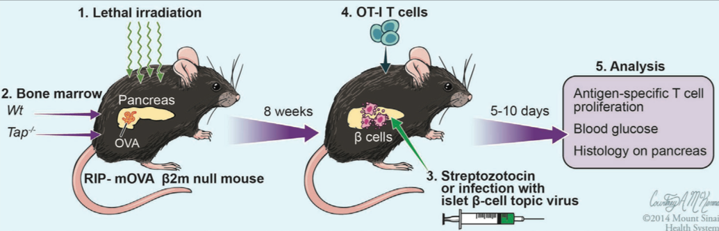
Depiction of an experimental scheme for manipulation of a mouse model for a grant application. Collaboration with Julie Blander.
In general, what do medical illustrators do?
Medical illustrators create graphics for all types of uses; from high school textbooks, to peer-reviewed journals, to visual evidence in malpractice cases. Most medical illustrators have a background in both the sciences and the arts, and the vast majority earn a specialized graduate degree in medical illustration. Though the field is called “medical” illustration, we in fact create images for all types of scientific fields, from molecular biology, to comparative anatomy, to genetics, and beyond.
How do you create your illustrations? Do you draw them by hand or use a computer?
Historically, medical illustrations were created using pencil, pen and ink, watercolor, and airbrush. In the past 15 years, the field has shifted to almost 100% digitally-created artwork. Common softwares include Adobe Photoshop, Adobe Illustrator, Pixological ZBrush, Maya, and Cinema 4D. Many illustrators are also animators, creating motion graphics for professional presentations, pharmaceutical marketing, and patient education.But, even in this digital world, most illustrations start with a pencil sketch, which is then scanned into the computer. Digital media is used to complete the work. We use Wacom tablets and pressure sensitive monitors to create our artwork- we don’t draw using a mouse!
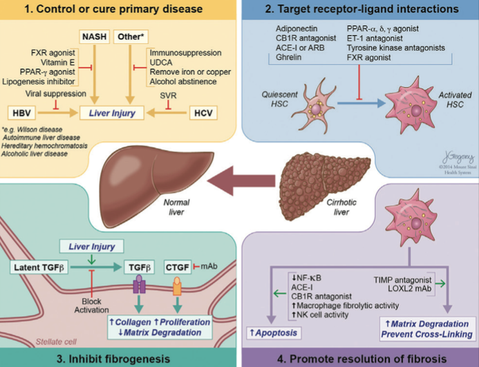
Mechanisms by which antifibrotic therapies may lead to fibrosis regression in the liver. This illustration accompanies the article “Pathology of liver fibrosis: a translational success story” by Youngmin A Lee, Michael C Wallace, and Scott L Friedman (Gut, in press).
How long have you worked at Mount Sinai?
We joined Mount Sinai through the merger with Continuum Health Partners in October 2013. We look forward to working with the range of research and clinical departments within the new health system!
What kind of projects do you work on?
A typical project is a set of illustrations for a peer-reviewed journal paper on anything from surgical techniques to cellular processes. We also create images for grant proposals, in-classroom lectures in the medical and graduate schools, and professional presentations at national conferences. See below for two examples of recent work.
How long does it take for you to complete a project?
This can vary quite a bit. A simple diagram can be completed in about a week, whereas a complicated, multi-step set of images can take 1-2 months. We provide better estimates once we know the details of a project.
Is there a fee for your services?
At this time, there is no charge.
How do I get in contact with you when I have an illustration project?
The easiest way to get ahold of us is by phone or email (see contact information above). We split our projects based onourschedulesandareas of interest, so feel free to contact either or both of us with a project request. We often complete projects communicating solely via email or phone, but if you’d like to meet in person, the Academic Medical Illustration office is located in the Levy Library, in room 11-10.